Contest on HEp-2 Cells Classification
Hosted by the 21th International Conference on Pattern Recognition
cfp
background
description
dataset
organizers
important dates
entry submission
results
Description of the dataset to be used
In the following we provide few notes about the dataset that will be used for the competition. Note that the dataset will be made available only to researchers registered to the contest. Registrations will start on November 15, 2011.
Acquisition and manual labeling procedure
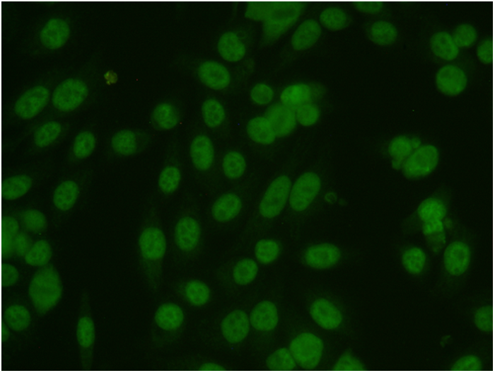
HEp-2 images were acquired by means of a fluorescence microscope (40-fold magnification) coupled with a 50Wmercury vapor lamp and with a digital camera (SLIM system by Das srl). The camera has a CCD with square pixel of 6.45 m. The images have a resolution of 1388x1038 pixels, a color depth of 24 bits and they are stored in BMP format (see the following picture for an example).
Specialists manually segmented and annotated each cell at a workstation monitor, and reported data on fluorescence intensity and staining pattern. Firstly, a biomedical engineer manually segmented the cells by the use of a tablet PC. Subsequently, each image was verified and annotated by a medical doctor specialized in immunology. The image database is annotated with the following information:
- Image main pattern and intensity;
- Cell seed points;
- Cell pattern, which can be one of: homogeneous, fine speckled, coarse speckled, nucleolar, cytoplasmatic or centromere.
Composition
The dataset is constituted by 28 images, which, as a whole, contain 1457 cells, so divided among the different patterns:
- Number of cells: 1457
- Centromere: 388
- Coarse Speckled: 239
- Cytoplasmatic: 128
- Fine Speckled: 225
- Homogeneous: 345
- Nucleolar: 257
Acknowledgments
The dataset used for the competition is one of the outcomes the research project "Classification of Immunofluorescence Images for the Diagnosis of Autoimmune Diseases" supported by "Regione Campania (Italy)".