Contest on HEp-2 Cells Classification
Hosted by the 21th International Conference on Pattern Recognition
cfp
background
description
dataset
organizers
important dates
entry submission
results
Background
Indirect ImmunoFluorescence (IIF) is considered a powerful, sensitive, and comprehensive test for antinuclear autoantibodies (ANAs) analysis. Furthermore, it is the most effective and widely-used diagnostic screening assay able to detect in a timely manner immune pathologies whose incidence has been constantly growing in the last few years.
IIF slides are examined at the fluorescence microscope, and their diagnosis requires both the estimation of the fluorescence intensity and the description of the staining pattern. The fluorescence intensity is scored semi-quantitatively with respect to both positive and negative controls contained in each slide. The staining pattern suggests the localization of reactive nuclear antigens and may help clinicians in differential diagnosis.
However, the IIF method has some disadvantages. The major ones are: the low level of standardization, the interobserver variability, which limits the reproducibility of IIF readings; the lack of resources and adequately trained personnel; the photobleaching effect, which bleaches significantly the tissues in a few seconds. Such drawbacks affect the diagnosis repeatability, therefore limiting the procedure reliability. Indeed, humans are limited in their ability to detect and diagnose a disease during image interpretation due to their non-systematic search patterns and to the presence of noise. In addition, the vast amount of image data that is generated makes the detection of potential disease a burdensome task and may cause oversight errors. Another problem is that similar characteristics of some abnormal and normal structures may cause interpretational errors. These problems result in a intra-laboratory variability estimated in the literature equal to 7-10%.
Automation may offer a solution to the growing demand of diagnostic tests for systemic autoimmune diseases, as in other areas of medicine. Being able to automatically determine the presence of autoantibodies in IIF would enable easier, faster and more reliable tests. Hence, an evident medical demand is the development of a Computer-Aided Diagnosis (CAD) system, which may support the physician's decision and overcome current method limitations. Indeed CAD methods have definitely been proven effective in other contexts as they:
- allow to perform a pre-selection of the cases to be examined, enabling the physician to focus his/her attention only on relevant cases, making it easier to carry out mass screening campaigns,
- serve as a second reader, thus augmenting the physician's capabilities and reducing errors,
- aid the physician while he/she carries out the diagnosis,
- work as a tool for training and education of specialized medical personnel.
Besides providing image acquisition and traditional image post-processing tools, the main functionality of a CAD regards the automatic classification of the images. The analysis of the literature in the field of ANAs detection reveals that a comprehensive CAD system in IIF is not available yet, while the use of digital images in IIF have been recently validated and recent research on partial CAD system can be found in the literature.
The flow of IIF diagnostic procedure consists of four main steps, namely image acquisition, mitosis detection, fluorescence intensity classification and staining pattern recognition. Among such steps, the last one is very challenging as well as important since several different patterns may be observed which match with different autoimmune diseases. In the literature, staining patterns are classified into one of the following groups:
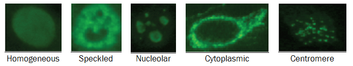
- homogeneous : diffuse staining of the interphase nuclei and staining of the chromatin of mitotic cells;
- speckled : a fine or coarse granular nuclear staining of the interphase cell nuclei;
- nucleolar : large coarse speckled staining within the nucleus, less than six in number per cell;
- cytoplasmic : fine fluorescent fibres running the length of the cell; it is frequently associated with other autoantibodies to give a mixed pattern;
- centromere : several discrete speckles (~40-60) distributed throughout the interphase nuclei and characteristically found in the condensed nuclear chromatin during mitosis as a bar of closely associated speckles.